Simple Compact Bone Diagram / 33 2c Connective Tissues Bone Adipose And Blood Biology Libretexts - Each cell appears to be isolated from other cells, but in reality are connected to neighboring cells by thin cellular extensions that pass through tiny channels in the solid matrix (see picture on left of.
Simple Compact Bone Diagram / 33 2c Connective Tissues Bone Adipose And Blood Biology Libretexts - Each cell appears to be isolated from other cells, but in reality are connected to neighboring cells by thin cellular extensions that pass through tiny channels in the solid matrix (see picture on left of.. The compact bones form the hard exterior of the bones, whereas the spongy bones have several pores that are filled with nerves and blood vessels. Microscopic anatomy bones medical massage physiology pediatric nursing medical knowledge medical anatomy anatomy tutorial structure of bone. Compact bone, also called cortical bone, is the hard, stiff, smooth, thin, white bone tissue that surrounds all bones in the human body. Create flashcards for free and quiz yourself with an interactive flipper. Related posts of compact bone diagram labeled anatomy of rib cage.
Long bones such as the femur contain two distinct morphological types of bone: Learn about the process of bone formation. Under the periosteum is a thin layer of compact bone (often called cortical bone). It makes up the outer cortex of all bones and is in immediate contact with the periosteum. The remainder is cancellous bone, which has a spongelike appearance with numerous large spaces and is found in the.
Simple Bone Diagram By Tessa Arnett Teachers Pay Teachers from ecdn.teacherspayteachers.com As seen in the image below, compact bone forms the cortex, or hard outer shell of most bones in the body. (b) in this micrograph of the osteon, you can clearly see the concentric lamellae and central canals. It makes up the outer cortex of all bones and is in immediate contact with the periosteum. They are roughly cylindrical, and about 0.2mm wide and a few millimeters long. Anatomy of rib cage 12 photos of the anatomy of rib cage anatomical rib cage necklace, anatomy and physiology of rib cage, anatomy of human rib cage, anatomy of rib cage area, human anatomy rib cage muscles, human anatomy, anatomical rib cage necklace, anatomy and physiology of rib cage, anatomy of human rib cage, anatomy … The remainder of the bone is formed by cancellous or spongy bone. Long bones such as the femur contain two distinct morphological types of bone: Diagram of a typical long bone:
The remainder of the bone is formed by cancellous or spongy bone.
The diagram above shows a longitudinal view of an osteon. Compact bone is made of a matrix of hard mineral salts reinforced with tough collagen fibers. Some, mostly older, compact bone is remodelled to form these haversian systems (or osteons). The remainder of the bone is formed by cancellous or spongy bone. Ossification begins about the third month of fetal life in humans and is completed by late adolescence. Start studying compact bone structure. Learn vocabulary, terms, and more with flashcards, games, and other study tools. Long bones such as the femur contain two distinct morphological types of bone: Microscopic anatomy bones medical massage physiology pediatric nursing medical knowledge medical anatomy anatomy tutorial structure of bone. Compact bone, also called cortical bone, dense bone in which the bony matrix is solidly filled with organic ground substance and inorganic salts, leaving only tiny spaces (lacunae) that contain the osteocytes, or bone cells.compact bone makes up 80 percent of the human skeleton; Diagram of distinct morphological types of bone. The compact bones form the hard exterior of the bones, whereas the spongy bones have several pores that are filled with nerves and blood vessels. Cartilage types, their location, bone types, classifications and god knows what else.
Some, mostly older, compact bone is remodelled to form these haversian systems (or osteons). The remainder is cancellous bone, which has a spongelike appearance with numerous large spaces and is found in the. The remainder of the bone is formed by cancellous or spongy bone. Compact bone, also called cortical bone, is the hard, stiff, smooth, thin, white bone tissue that surrounds all bones in the human body. Deep to the compact bone layer is a region of spongy bone where the bone tissue grows in thin columns called.
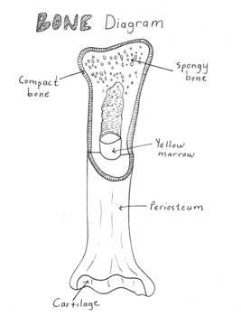
6 3 Bone Structure Anatomy Physiology from open.oregonstate.education In compact bone, these cells are embedded within the solid calcium phosphate matrix of solid bone. Create flashcards for free and quiz yourself with an interactive flipper. Simple bone diagrams to help students identify and label compact bone, spongy bone, yellow marrow, periosteum, and cartilage. About press copyright contact us creators advertise developers terms privacy policy & safety how youtube works test new features press copyright contact us creators. They are roughly cylindrical, and about 0.2mm wide and a few millimeters long. Shows compact (cortical) and cancellous (spongy) bone. Many tiny cells called osteocytes live in small spaces in the matrix and help to maintain the strength and integrity of the compact bone. Learn about the process of bone formation.
(b) in this micrograph of the osteon, you can clearly see the concentric lamellae and central canals.
Compact bone, as opposed to spongy bone, is made of cylindrical units, called osteons, that are tightly formed together. Over several more weeks or months, compact bone replaces spongy bone at the outer margins of the fracture and the bone is remodeled in response to strain (figure 6.5.2d). About press copyright contact us creators advertise developers terms privacy policy & safety how youtube works test new features press copyright contact us creators. The process takes two general forms, one for compact bone and the other for cancellous bone. The diagram above shows a longitudinal view of an osteon. Create flashcards for free and quiz yourself with an interactive flipper. In compact bone, these cells are embedded within the solid calcium phosphate matrix of solid bone. The main type of bone cell is the osteocyte (bone cell, shown as purple in the diagram). Diagram of distinct morphological types of bone. Diagram of a typical long bone: Bones are hard structures because of the calcium that builds up in the matrix, but they retain a slight amount of elasticity due to the fibers. Each cell appears to be isolated from other cells, but in reality are connected to neighboring cells by thin cellular extensions that pass through tiny channels in the solid matrix (see picture on left of. This provides the bones strength and consists of tightly stacked layers of bone which appear to form a solid section.
Article by jennifer smith owens. Microscopic anatomy bones medical massage physiology pediatric nursing medical knowledge medical anatomy anatomy tutorial structure of bone. Create flashcards for free and quiz yourself with an interactive flipper. Bones are hard structures because of the calcium that builds up in the matrix, but they retain a slight amount of elasticity due to the fibers. About press copyright contact us creators advertise developers terms privacy policy & safety how youtube works test new features press copyright contact us creators.
Structure Of Bones Biology For Majors Ii from s3-us-west-2.amazonaws.com Diagram of a typical long bone: Shows compact (cortical) and cancellous (spongy) bone. Bone formation, process by which new bone is produced. Ossification begins about the third month of fetal life in humans and is completed by late adolescence. Simple bone diagrams to help students identify and label compact bone, spongy bone, yellow marrow, periosteum, and cartilage. Each cell appears to be isolated from other cells, but in reality are connected to neighboring cells by thin cellular extensions that pass through tiny channels in the solid matrix (see picture on left of. Compact bone is the denser, stronger of the two types of osseous tissue (figure 6.3.6). As seen in the image below, compact bone forms the cortex, or hard outer shell of most bones in the body.
Cancellous or trabecular (spongy) bone;
About press copyright contact us creators advertise developers terms privacy policy & safety how youtube works test new features press copyright contact us creators. Each cell appears to be isolated from other cells, but in reality are connected to neighboring cells by thin cellular extensions that pass through tiny channels in the solid matrix (see picture on left of. Your bones contain a matrix, osteoblasts, osteoclasts, osteocytes, and collagen fibers. Bones are hard structures because of the calcium that builds up in the matrix, but they retain a slight amount of elasticity due to the fibers. Under the periosteum is a thin layer of compact bone (often called cortical bone). Compact bone, also called cortical bone, is the hard, stiff, smooth, thin, white bone tissue that surrounds all bones in the human body. Some, mostly older, compact bone is remodelled to form these haversian systems (or osteons). About press copyright contact us creators advertise developers terms privacy policy & safety how youtube works test new features press copyright contact us creators. This provides the bones strength and consists of tightly stacked layers of bone which appear to form a solid section. Compact bone is made of a matrix of hard mineral salts reinforced with tough collagen fibers. (b) in this micrograph of the osteon, you can clearly see the concentric lamellae and central canals. As seen in the image below, compact bone forms the cortex, or hard outer shell of most bones in the body. Over several more weeks or months, compact bone replaces spongy bone at the outer margins of the fracture and the bone is remodeled in response to strain (figure 6.5.2d).
Some, mostly older, compact bone is remodelled to form these haversian systems (or osteons) compact bone diagram. Deep to the compact bone layer is a region of spongy bone where the bone tissue grows in thin columns called.


0 Komentar